- Amputation of digits
- Partial foot amputation (Chopart, Lisfranc)
- Ankle disarticulation (Syme, Pyrogoff)
- Below-knee amputation (transtibial)
- Knee-bearing amputation (knee disarticulation)
- Above knee amputation (transfemoral)
- Van-ness rotation/rotationplasty (Foot being turned around and reattached to allow the ankle joint to be used as a knee.)
- Hip disarticulation
- Hemipelvectomy
Sunday, August 31, 2008
Types of Leg Amputations
Eisenmenger's Syndrome
Description:
It is predominantly right-to-left shunt or reversal of left-to-right shunt, which usually resuts from severe pulmonary vascular obstruction.
Complications:1. Stroke
2. Hemoptysis
3. Hyperviscosity
4. Hemostatic abnormalities.
5. Thrombocytopenia
6. Prolonged bleeding, prothrombin or partial thromboplastin times.
7. Vitamin K-dependent clotting factors deficiencies.
8. Abnormal fibrinolysis.
9. Cholelithiasis
10. Hypertrophic osteoarthropathy
11. Hyperuricemia and goutrenal dysfunction.
12. Sudden death.
Clinical Features:
Symptoms : dyspnea on exertion, chest pain, syncope, hemoptysis, angina.
Signs : cyanosis (constant or exercise), ascites, pedal edema, clubbing.
Investigations:
1. Blood tests :
a) 100% O2 does not correct arterial desaturation.
b) polycythemia (Hb > 20 g/dl common)
2. Imaging studies : echocardiography, Doppler, hemogram, catheterization
ECG shows right ventricular hypertrophy changes.
Treatment:
Surgery is indicated if medical treatment does not improve the oxygen saturation levels.
Phlebotomy may be necessary.
Saturday, August 30, 2008
Acute otitis media
Causes:
1. Viruses - most commonly rhinovirus, RSV and coronavirus.
2. Bacteria - Streptococcus pneumoniae, Moraxella (Branhamella) catarrhalis, H. influenzae and Streptococcus pyogenes (rare).
Pathogenesis:
Eustachian tube is port of entry for middle-ear pathogens from nasopharynx, but also is primary route of clearing middle ear secretions. This causes inflammatory edema of nasopharynx. Following this exudative and transudative fluid collect in middle ear, which allows for overgrowth of nasopharyngeal bacteria in middle ear. This suppuration may lead to spontaneous rupture, usually anterior-inferior quadrant following which discharge is seen, which is initially serosanguineous and later becomes mucopurulent.
Complications:
1. Hearing loss
2. Mastoiditis
3. Meningitis
Clinical Features:
1. Redness with bulging of tympanic membrane.
2. Mobility may be impaired.
Treatment:
A: Medical
1. Analgesics (ibuprofen and acetaminophen)
2. Antibiotics (amoxicillin is drug of choice)
3. Decongestants and/or Antihistamines (not in children)
B: Surgical
1. Tympanostomy tubes reduce recurrences of otitis media when middle ear effusion present.
2. Myringotomy to drain the pus.
3. Adenoidectomy is not very effective.
Thursday, August 28, 2008
Astigmatism
Definition:
It is the condition in which an optical system has different foci for rays that propagate in two perpendicular planes. If an optical system with astigmatism is used to form an image of a cross, the vertical and horizontal lines will be in sharp focus at two different distances.
Types:
A: Based on axis of the principal meridians
1. Regular
- With the rule
- Against the rule
- Oblique
- Bioblique
2. Irregular
B. Based on focus of the principal meridians
1. Simple
- Myopic
- Hypermetropic
2. Compund
- Myopic
- Hypermetropic
3. Mixed
Symptoms:
1. Blurring of vision
2. Squinting
3. Asthenopic symptoms
4. Headache
Treatment:
Astigmatism may be corrected with eyeglasses, contact lenses, or refractive surgery.
Spherical lenses correction along with a cylindrical lens for correction of the residual error is used.
Wednesday, August 27, 2008
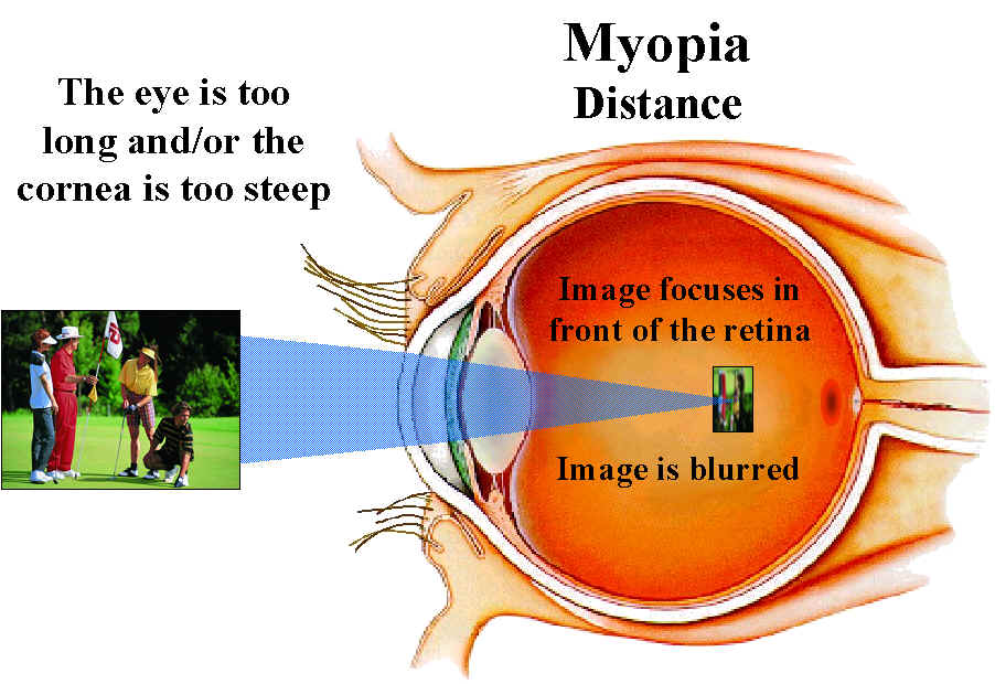
Myopia
Definition:
It is that dioptric condition of the eye in which parallel rays of light are brought to focus at a point in front of the retina, in an eye with accomodation at rest.
 Classification (based on etiology):
1. Axial myopia
2. Refractive myopia
It is that dioptric condition of the eye in which parallel rays of light are brought to focus at a point in front of the retina, in an eye with accomodation at rest.
 Classification (based on etiology):
1. Axial myopia
2. Refractive myopia
- Curvature myopia
- Index myopia
Treatment:
- Eyeglasses
- Contact Lenses
- Refractive Surgery
Sunday, August 24, 2008
Cataract - Diagnosis
- Slit-lamp biomicroscopy : To detect location and density of opacity within lens.
- Potential visual acuity with removal of cataract can be estimated with potential acuity meter (PAM) or laser interferometer.
- B-scan ultrasound can be used to detect retinal detachment or tumors if severe cataract prevents retinal examination.
- Glare testing and contrast sensitivity testing can quantitatively estimate functional impact of cataract on vision.
Cataract (contd..)
Risk Factors:
- Inhaled steroids
- Tobacco use
- Exposure of UV-B light
- Allopurinol
- Trabeculectomy
- Diabetes
- Psychotropic medications (antipsychotics, antidepressants, lithium, anticonvulsants, benzodiazepines)
- Alcohol consumption
- Obesity
- Medications for hypertension, and angina
Cataract - Classification (contd..)
Classification based on location of the opacity:
1. Anterior cortical cataract
2. Anterior polar cataract
3. Anterior subcapsular cataract
4. Posterior cortical cataract
5. Posterior polar cataract
6. Posterior subcapsular cataract (PSC)
7. Nuclear cataract
Grading:
1. Anterior cortical cataract
2. Anterior polar cataract
3. Anterior subcapsular cataract
4. Posterior cortical cataract
5. Posterior polar cataract
6. Posterior subcapsular cataract (PSC)
7. Nuclear cataract
Grading:
- Grey
- Yellow
- Amber
- Brown/Black
Saturday, August 23, 2008
Cataract - Classification
Etiological Classification
1. Age-related cataract
1. Age-related cataract
- Immature Senile Cataract (IMSC)
- Cataract (MSC)
- Senile Cataract (HMSC)
2. Congenital cataract
- Sutural cataract
- Lamellar cataract
- Zonular cataract
- Total cataract
3. Secondary cataract
- Drug-induced cataract (e.g. Corticosteroids)
4. Traumatic cataract
- Blunt trauma
- Penetrating trauma
Thursday, August 21, 2008
Cataract
Definition:

A cataract is a clouding that develops in the crystalline lens of the eye or in its envelope, varying in degree from slight to complete opacity and obstructing the passage of light.
Tuesday, August 19, 2008
Pterygium
Definition:
Pterygium is a raised, wedge-shaped growth of the conjunctiva. It is most common among those who live in tropical climates or spend a lot of time in the sun.

Pathology:
Pterygium in the conjunctiva is characterized by elastotic degeneration of collagen and fibrovascular proliferation. It has an advancing portion called the head of the pterygium, which is connected to the main body of the pterygium by the neck. Sometimes a line of iron deposition can be seen adjacent to the head of the pterygium called Stocker's line. The location of the line can give an indication of the pattern of growth.
Anatomically, the pterygium is comprised of several segments:
Symptoms of pterygium include
The majority of pterygia are inactive and can be treated with topical drops. Decongestant eye drops to make the eye appear less red and artificial tear drops to make the eye more comfortable when the pterygium flares up, can be used. If these are sufficient to maintain comfort and cosmesis then surgery is not indicated.
If this fails:
Pterygium is a raised, wedge-shaped growth of the conjunctiva. It is most common among those who live in tropical climates or spend a lot of time in the sun.
Pathology:
Pterygium in the conjunctiva is characterized by elastotic degeneration of collagen and fibrovascular proliferation. It has an advancing portion called the head of the pterygium, which is connected to the main body of the pterygium by the neck. Sometimes a line of iron deposition can be seen adjacent to the head of the pterygium called Stocker's line. The location of the line can give an indication of the pattern of growth.
Anatomically, the pterygium is comprised of several segments:
- Fuchs' Patches (minute gray blemishes that disperse near the pterygium head).
- Stocker's Line (a brownish line composed of iron deposits).
- Hood (fibrous nonvascular portion of the pterygium).
- Head (apex of the pterygium, typically raised and highly vascular).
- Body (fleshy elevated portion congested with tortuous vessels).
- Superior Edge (upper edge of the triangular or wing shaped portion of the pterygium).
- Inferior Edge (lower edge of the triangular or wing shaped portion of the ptyerygium).
Symptoms of pterygium include
- Persistent redness
- Inflammation
- Foreign body sensation
- Dry and itchy eyes.
- In advanced cases the pterygium can affect vision as it invades the cornea with the potential of induced astigmatism and corneal scarring.
The majority of pterygia are inactive and can be treated with topical drops. Decongestant eye drops to make the eye appear less red and artificial tear drops to make the eye more comfortable when the pterygium flares up, can be used. If these are sufficient to maintain comfort and cosmesis then surgery is not indicated.
If this fails:
- The 90 Sr plaque is a concave metal disc about 1-1.5cm in diameter which is hollow and filled with an insoluble strontium salt. The side placed on the eye is a very thin and delicate silver film that will contain the strontium but allow the beta particles to escape. The dose of radiation to the conjunctiva is controlled by the time that the plaque is left in contact with the surface. The integrity of the plaque surfaces is paramount to prevent exposure to patients and so is wipe tested to see if radioactive matter is escaping.
- Conjunctival auto-grafting is a surgical technique that is effective and safe procedure for pterygium removal. When the pterygium is removed, the conjunctiva is also extracted, which is replaced from another peice of conjunctiva from the same patient.
Sunday, August 17, 2008
Hordeolum
Definition:
A common staphyloccal infection of the lid glands, essentially an abscess, with pus formation.
Classification:
Two types
The patient complains of redness, swelling, and pain in the eyelid and swelling which is:
A common staphyloccal infection of the lid glands, essentially an abscess, with pus formation.
Classification:
Two types
- Internal hordeolum - relatively large, affecting the meibomian glands. May point towards the skin or towards the conjunctivae.
- External hordeolum - also known as a stye. It is smaller and more superficial. It is an infection of the glands of Moll or Zeiss and it always points toward the skin side of the lid margin.


The patient complains of redness, swelling, and pain in the eyelid and swelling which is:
- At the base of an eyelash (external hordeolum)
- Deep within the lid (internal hordeolum)
Treatment:
Both types of hordeola are treated with warm compresses for 10-15 minutes 3-4 times a day. If the condition does not improve within 48 hours, incision and drainage of the pus is indicated. Antibacterial ophthalmic ointment is also helpful.
Wednesday, August 13, 2008
Chalazion
Description:
A chalazion, also known as a meibomian gland lipogranuloma, is a cyst in the eyelid that is caused by inflammation of a blocked meibomian gland, usually on the upper eyelid.

Signs and symptoms:
- Swelling on the eyelid
- Eyelid tenderness
- Sensitivity to light
- Increased tearing
- Heaviness of the eyelid
Treatment:
- The primary treatment is application of warm compresses for 10 to 20 minutes at least 4 times a day. This may soften the hardened oils blocking the duct and promote drainage and healing.
- Topical antibiotic eye drops or ointment.
- Corticosteroid injections
- Surgery under local anesthesia.
Complications:
- A large chalazion can cause astigmatism due to pressure on the cornea.
- Hypopigmentation may occur with corticosteroid injection.
- Sebaceous cell carcinoma (recurring chalazion).
Monday, August 11, 2008
Legg-Calvé-Perthes Syndrome (Perthes Disease)
Definition:
Legg-Calvé-Perthes syndrome is a degenerative disease of the hip joint, where a loss of bone mass leads to some degree of collapse of the hip joint, that is, to deformity of the ball of the femur and the surface of the hip socket. The disease is typically found in young children and small dogs, and it can lead to osteoarthritis in adults. Perthes can also sometimes continue into adulthood.
It is the idiopathic avascular osteonecrosis of the capital femoral epiphysis of the femoral head. It is caused by an interruption to the blood supply of the head of the femur close to the hip joint. It is equivalent to adult avascular necrosis.
Cause:
The direct cause is a reduction in blood flow to the joint, though what causes this is unknown. It is thought that the artery of the ligamentum teres femoris closes too early, not allowing time for the medial circumflex femoral artery to take over.[citation needed]Genetics does not appear to be a determining factor, though it may be involved. When the disease is genetic in origin, it typically runs along the male line. Some evidence suggests that parental smoking may be a factor, though this is not yet proven, or more recently that a deficiency of some blood factors used to disperse blood clots may lead to blockages in the vessels supplying the joint, but that, too, has not been proven.
Symptoms:
Symptoms are hip or groin pain, exacerbated by hip/leg movement. There is a reduced range of motion at the hip joint and a painful or antalgic gait. There may be atrophy of thigh muscles from disuse and an inequality of leg length. In some cases, some activity can cause severe irritation or inflammation of the damaged area including standing, walking, running, kneeling, or stooping repeatedly for an extended period of time.
Signs:
The first signs are complaints of soreness from the child, particularly when tired. The pain is usually in the hip, referred to the knee. It is predominantly a disease of boys (4:1 ratio). Whereas Perthes is generally diagnosed between 5 and 12 years of age. Typically the disease is only seen in one hip.
Treatment:
The goal of treatment is to avoid severe degenerative arthritis. Orthopedic assessment is crucial. Younger children have a better prognosis than older children. Currently, there are studies conducted on bisphosphonates for treatment of Perthes. Analgesic medication may be given as necessary.
Subscribe to:
Comments (Atom)