Saturday, December 15, 2007

Mesothelioma Update

Malignant mesothelioma is a rare cancer that occurs in the thin layer of tissue that covers the majority of your internal organs (mesothelium). Doctors divide mesothelioma into different categories based on what part of the mesothelium is affected by cancer. Mesothelioma that occurs in the tissue that surrounds the lung (pleura) is called pleural mesothelioma and is the most common form. Mesothelioma that occurs in the tissue in your abdomen (peritoneum) is called peritoneal mesothelioma and accounts for 10 percent to 20 percent of all mesotheliomas. In rare cases, mesothelioma can also occur in the lining around the heart (pericardium) and in the lining around the testicles (tunica vaginalis).

Friday, December 14, 2007

Osteomyelitis

Osteomyelitis is an infection of bone or bone marrow, usually caused by pyogenic bacteria or mycobacteria. It can be usefully subclassifed on the basis of the causative organism, the route, duration and anatomic location of the infection.


Wednesday, December 12, 2007

Deep Vein Thrombosis (DVT)

Deep-vein thrombosis (also known as deep-venous thrombosis or DVT and colloquially as economy class syndrome) is the formation of a blood clot ("thrombus") in a deep vein. It commonly affects the leg veins, such as the femoral vein or the popliteal vein or the deep veins of the pelvis. Occasionally the veins of the arm are affected (known as Paget-Schrötter disease). Thrombophlebitis is the more general class of pathologies of this kind. There is a significant risk of the thrombus embolizing and traveling to the lungs causing a pulmonary embolism.

Sunday, December 9, 2007

Disseminated Intravascular Coagulation

Disseminated intravascular coagulation (DIC), also called consumptive coagulopathy, is a pathological process in the body where the blood starts to coagulate throughout the whole body. This depletes the body of its platelets and coagulation factors, and there is a paradoxically increased risk of hemorrhage. It occurs in critically ill patients, especially those with Gram-negative sepsis (particularly meningococcal sepsis) and acute promyelocytic leukemia.

The treatment of underlying cause is a must and should be initiated immediately. Anticoagulants are only given when indicated (development of thrombotic renal complications) as patients with DIC are prone to bleeding. Platelets may be transfused if counts are very low, and fresh frozen plasma may be administered.
DIC results in lower fibrinogen (as it has all been converted to fibrin), and this can be tested for in the hospital lab. A more specific test is for "fibrin split products" (FSPs) or "fibrin degradation products" (FDPs) which are produced when fibrin undergoes degradation when blood clots are dissolved by fibrinolysis.
In some situations, infusion with antithrombin may be necessary. A new development is drotrecogin alfa, a recombinant activated protein C product. Activated Protein C (APC) deactivates clotting factors V and VIII, and the presumed mechanism of action of drotrecogin is the cessation of the intravascular coagulation. Due to its high cost, it is only used strictly on indication in intensive care patients.
The prognosis for those with DIC, depending on its cause, is often grim, leading the initials to be known colloquially as "death is coming".

Source : Wikipedia

Saturday, December 8, 2007

Bronchiectasis

Inrtoduction:

Bronchiectasis is a disease that causes localized, irreversible dilatation of part of the bronchial tree. Involved bronchi are dilated, inflamed, and easily collapsible, resulting in airflow obstruction and impaired clearance of secretions. Bronchiectasis is associated with a wide range of disorders, but it usually results from necrotizing bacterial infections, such as infections caused by the Staphylococcus or Klebsiella species or Bordetella pertussis.

Pathogenesis:
Dilation of the bronchial walls results in airflow obstruction and impaired clearance of secretions because the dilated areas disrupt normal air pressure in the bronchial tubes, causing sputum to pool inside the dilated areas instead of being pushed upward. The pooled sputum provides an environment conducive to the growth of infectious pathogens, and these areas of the lungs are thus very vulnerable to infection. The more infections that the lungs experience, the more damaged the lung tissue and alveoli become. When this happens, the bronchial tubes become more inelastic and dilated, creating a self-perpetuating cycle of further damage to the lungs.

Causes:
There are both congenital and acquired causes of bronchiectasis. Kartagener syndrome, which affects the mobility of cilia in the lungs, aids in the development of the disease. Another common genetic cause is cystic fibrosis, in which a small number of patients develop severe localized bronchiectasis. Young's syndrome, which is clinically similar to cystic fibrosis, is thought to significantly contribute to the development of bronchiectasis. This is due to the occurrence of chronic, sinopulmonary infections. Patients with alpha 1-antitrypsin deficiency have been found to be particularly susceptible to bronchiectasis, for unknown reasons. Other less-common congenital causes include primary immunodeficiencies, due to the weakened or nonexistent immune system response to severe, recurrent infections that commonly affect the lung.

Treatment:
Treatment of bronchiectasis is aimed at controlling infections and bronchial secretions, relieving airway obstruction, and preventing complications. This includes the prolonged usage of antibiotics to prevent detrimental infections, as well as eliminating accumulated fluid with postural drainage and chest physiotherapy. Surgery may also be used to treat localized bronchiectasis, removing obstructions that could cause progression of the disease.
Inhaled steroid therapy that is consistently adhered to can reduce sputum production and decrease airway constriction over a period of time, and help prevent progression of bronchiectasis. One commonly used therapy is beclometasone dipropionate, which also used in asthma treatment. Use of inhalers such as albuterol (salbutamol), fluticasone (Flovent/Flixotide) and ipratropium (Atrovent) may help reduce likelihood of infection by clearing the airways and decreasing inflammation.

Thursday, December 6, 2007

Mesothelioma

Mesothelioma is a form of cancer that is almost always caused by previous exposure to asbestos. In this disease, malignant cells develop in the mesothelium, a protective lining that covers most of the body's internal organs. Its most common site is the pleura (outer lining of the lungs and chest cavity), but it may also occur in the peritoneum (the lining of the abdominal cavity) or the pericardium (a sac that surrounds the heart).

Most people who develop mesothelioma have worked on jobs where they inhaled asbestos particles, or have been exposed to asbestos dust and fibre in other ways, such as by washing the clothes of a family member who worked with asbestos, or by home renovation using asbestos cement products. Unlike lung cancer, there is no association between mesothelioma and smoking.

Source : Wikipedia

Wednesday, December 5, 2007

Tuesday, December 4, 2007

Meckel's diverticulum

Introduction:
A Meckel's diverticulum, a true congenital diverticulum, is a small bulge in the small intestine present at birth. It is a vestigial remnant of the omphalomesenteric duct (also called the vitelline duct), and is the most frequent malformation of the gastrointestinal tract. It is present in approximately 2% of the population, with males more frequently experiencing symptoms.

Symptoms:
If symptoms do occur, they typically appear before the age of two.
The most common presenting symptom is painless rectal bleeding, followed by intestinal obstruction, volvulus and intussusception. Occasionally, Meckel's diverticulitis may present with all the features of acute appendicitis. Also, severe pain in the upper abdomen is experienced by the patient along with bloating of the stomach region. At times, the symptoms are so painful such that they may cause sleepless nights with extreme pain in the abdominal area.

Treatment:
Treatment is surgical, consisting of a resection of the affected portion of the bowel.

Whipple's disease

Introduction:
Whipple's disease is a rare, systemic infectious disease caused by the bacterium Tropheryma whipplei. First described by George Hoyt Whipple in 1907 and commonly considered a gastrointestinal disorder, Whipple's disease primarily causes malabsorption but may affect any part of the body including the heart, lungs, brain, joints, and eyes. Weight loss, diarrhea, joint pain, and arthritis are common presenting symptoms, but the presentation can be highly variable.


This micrograph shows the typical findings of enlarged villus with abundant macrophages in the in small intestine.


Symptoms:
  • Arthritis or arthralgia
  • malabsorption
  • intestinal lipodystrophy (accumulation of fatty deposits in lymph nodes of the intestine)
  • lymphadenopathy
  • abdominal pain
  • diarrhea
  • fever
  • melanoderma
  • Neurological symptoms

Treatment:

Treatment is with penicillin, ampicillin, tetracycline or co-trimoxazole for 1 -2 years.

Sunday, December 2, 2007

Crohn's Disease

Introduction:
Crohn's disease (also known as regional enteritis) is a chronic, episodic, inflammatory condition of the gastrointestinal tract characterized by transmural inflammation (affecting the entire wall of the involved bowel) and skip lesions (areas of inflammation with areas of normal lining between). Crohn's disease is a type of inflammatory bowel disease (IBD) and can affect any part of the gastrointestinal tract from mouth to anus; as a result, the symptoms of Crohn's disease vary among afflicted individuals. The main gastrointestinal symptoms are abdominal pain, diarrhea (which may be bloody) or constipation, vomiting and weight loss. Crohn's disease can also cause complications outside of the gastrointestinal tract such as skin rashes, arthritis, and inflammation of the eye.


Symptoms:
Gastrointestinal symptoms:
Abdominal pain may be the initial symptom of Crohn's disease. The pain is commonly cramp-like and may be relieved by defecation. It is often accompanied by diarrhea, which may or may not be bloody, though constipation is not uncommon especially in those who have had surgery. The nature of the diarrhea in Crohn's disease depends on the part of the small intestine or colon that is involved. Ileitis typically results in large-volume watery feces. Colitis may result in a smaller volume of feces of higher frequency. Fecal consistency may range from solid to watery. In severe cases, an individual may have more than 20 bowel movements per day and may need to awaken at night to defecate. Visible bleeding in the feces is less common in Crohn's disease than in ulcerative colitis, but may be seen in the setting of Crohn's colitis. Bloody bowel movements are typically intermittent, and may be bright or dark red in colour. In the setting of severe Crohn's colitis, bleeding may be copious. Flatus and bloating may also add to the intestinal discomfort.


Systemic symptoms:
Crohn's disease, like many other chronic, inflammatory diseases, can cause a variety of systemic symptoms. Among children, growth failure is common. Many children are first diagnosed with Crohn's disease based on inability to maintain growth. As Crohn's disease may manifest at the time of the growth spurt in puberty, up to 30% of children with Crohn's disease may have retardation of growth. Fever may also be present, though fevers greater than 38.5 ˚C (101.3 ˚F) are uncommon unless there is a complication such as an abscess. Among older individuals, Crohn's disease may manifest as weight loss. This is usually related to decreased food intake, since individuals with intestinal symptoms from Crohn's disease often feel better when they do not eat and might lose their appetite. People with extensive small intestine disease may also have malabsorption of carbohydrates or lipids, which can further exacerbate weight loss.

Treatment:

Treatment is only needed for people exhibiting symptoms. The therapeutic approach to Crohn's disease is sequential: to treat acute disease and then to maintain remission. Treatment initially involves the use of medications to treat any infection and to reduce inflammation. This usually involves the use of aminosalicylate anti-inflammatory drugs and corticosteroids, and may include antibiotics.
Once remission is induced, the goal of treatment becomes maintaining remission and avoiding flares. Because of side-effects, the prolonged use of corticosteroids must be avoided. Although some people are able to maintain remission with aminosalicylates alone, many require immunosuppressive drugs.

Surgery may be required for complications such as obstructions, fistulas and/or abscesses, or if the disease does not respond to drugs within a reasonable time. For patients with an obstruction due to a stricture, two options for treatment are strictureplasty and resection of that portion of bowel. According to a retrospective review at the Cleveland Clinic, there is no statistical significance between strictureplasty alone versus strictureplasty and resection specifically in cases of duodenal involvement. In these cases, re-operation rates were 31% and 27%, respectively, indicating that strictureplasty is a safe and effective treatment for selected patients with duodenal involvement.


Source : Wikipedia

Saturday, December 1, 2007

Celiac Sprue

Introduction:
It is a malabsorption syndrome related to immune reaction to gluten in diet, also called nontropical sprue, celiac disease and gluten sensitive enteropathy (GSE).

Causes:
  • Toxic effect of gluten/gliadin (gliadin is a glycoprotein component of gluten)
  • immune reaction (antibody to gluten/gliadin)
  • adenovirus type 12
  • may be autosomal dominant with incomplete penetrance
Physical Findings:

In infants - growth retardation, failure to thrive and irritability. Most patients are thin, emaciated.
Microscopic Findings:

This is a micrograph of a typical celiac disease intestine.


Diagnosis:
There are several tests that can be used to assist in diagnosis. The level of symptoms may determine the order of the tests, but all tests lose their usefulness if the patient is already taking a gluten-free diet. Intestinal damage begins to heal within weeks of gluten being removed from the diet, and antibody levels decline over months. For those who have already started on a gluten-free diet, it may be necessary to perform a re-challenge with 10 g of gluten (four slices of bread) per day over 2–6 weeks before repeating the investigations. Those who experience severe symptoms (e.g. diarrhoea) earlier can be regarded as sufficiently challenged and can be tested earlier.


Prognosis:
Patient do recover and villi regenerate.
Celiac disease associated with increased mortality among patients with malabsorption symptoms.

Early Gastric Carcinoma

This is a gross specimen of an early gastric carcinoma (from Pathology Text).
It shows nodular mucosa, not very apparent from this surface view, but it is very much evident on cut section, showing involvement of mucosa and submucosa, but not the muscularis.

Hirschsprung's disease

Description:

Also called congenital megacolon. Its a disease in which there is congenital absence of parasympathetic innervation of distal intestine.


Pathogenesis:

Physical Findings:

Abdominal distention, occasionally loops of stool-filled bowel palpable, increased rectal sphincter tone, no stool in rectal vault or ampulla, classically explosion of watery stool on removal of finger.

Lab Diagnosis:

  • Abdominal x-ray
  • Barium enema
  • Rectal suction biopsy
  • No ganglion cells in submucosal plexus confirms diagnosis

Treatment:

Cured by surgical excision of affected segment.

Acute Gastritis

Description:
Focal damage to gastric mucosa. Also called acute erosive gastritis, gastric erosion, acute hemorrhagic gastritis, stress ulcers.

Causes:
  • Aspirin (NSAIDs)
  • Ethanol
  • Aalkali and acid
  • Ischemia
  • Steroids, cigarettes, pancreatic enzymes, bile acids
  • Severe stress such as sepsis, severe trauma and burns.

Physical Findings:

Often normal, GI bleeding, tachycardia, hypotension, pallor and upper abdominal tenderness.

Diagnosis:

  1. Endoscopy shows congestion, friability, superficial ulceration, petechiae, black hemorrhagic areas are superficial erosions
  2. Blood in gastric aspirate

Treatment:

  1. Antacids, H2 blockers, sucralfate, omeprazoleprochlorperazine for treating uncomplicated nausea and vomiting.
  2. For massive bleeding give vasopressin IV or intra-arteriorly to mesenteric artery; contraindicated in coronary artery disease; only temporary treatment.
  3. Near-total gastrectomy if uncontrolled bleeding

Thursday, November 29, 2007

Coming Up....

I shall be dealing with the diseases of the gut in the next few posts.
These include the following :
  1. Peptic Ulcer
  2. Gastritis
  3. Gastric Carcinoma
  4. Hirschsprung's disease
  5. Celiac sprue
  6. Crohn's disease
  7. Ulcerative colitis
  8. Paralytic Ileus
  9. Whipple disease
  10. Meckel's diverticulum

Poliomyelitis in India


A problem statement of Poliomyelitis in India.
This map shows the incidence of Wild Poliovirus in India.

Dengue Fever

Causes:
Caused by dengue virus (Flaviviridae, Arbovirus). 4 serotypes of this virus have been reported.
Characteristics of dengue virus: Icosahedral, envelope, internal matrix protein, 1 glycoprotein spike, single-stranded RNA.
It lives only in humans.
Transmitted by mosquito bite (Aedes aegypti mosquito, also Aedes albopictus and Aedes polynesiensis mosquitoes), typically during daytime.

Pathogenesis:
Incubation period 5-8 days (range 3-15 days)
It usually manifests as dengue hemorrhagic fever.
Caused by cross-reacting antibody at second infection and/or virulent virus and leakage of plasma through endothelial gaps.

Risk factors:
Mosquito season, travel to tropics and urban epidemics.
Endemic in Africa, Central and South America, Caribbean and southeast Asia.

Malaria : The Risk


Malaria incidence rates in different parts of the world.
Source : WHO World Malaria Report 2005

Tetanus

Introduction:
Tetanus is a medical condition that is characterized by a prolonged contraction of skeletal muscle fibers. The primary symptoms are caused by tetanospasmin, a neurotoxin produced by the Gram-positive, obligate anaerobic bacterium Clostridium tetani. Infection generally occurs through wound contamination, and often involves a cut or deep puncture wound. As the infection progresses, muscle spasms in the jaw develop hence the common name, lockjaw. This is followed by difficulty swallowing and general muscle stiffness and spasms in other parts of the body. Infection can be prevented by proper immunization and by post-exposure prophylaxis.

Types:
Local tetanus: It is an uncommon form of the disease, in which patients have persistent contraction of muscles in the same anatomic area as the injury. The contractions may persist for many weeks before gradually subsiding. Local tetanus is generally milder; only about 1% of cases are fatal, but it may precede the onset of generalized tetanus.
Cephalic tetanus :It is a rare form of the disease, occasionally occurring with otitis media (ear infections) in which C. tetani is present in the flora of the middle ear, or following injuries to the head. There is involvement of the cranial nerves, especially in the facial area.
Generalized tetanus: It is the most common type of tetanus, representing about 80% of cases. The generalized form usually presents with a descending pattern. The first sign is trismus or lockjaw, followed by stiffness of the neck, difficulty in swallowing, and rigidity of pectoral and calf muscles. Other symptoms include elevated temperature, sweating, elevated blood pressure, and episodic rapid heart rate. Spasms may occur frequently and last for several minutes. Spasms continue for 3–4 weeks and complete recovery may take months.
Neonatal tetanus: It is a form of generalized tetanus that occurs in newborn infants. It occurs in infants who have not acquired passive immunity because the mother has never been immunized. It usually occurs through infection of the unhealed umbilical stump, particularly when the stump is cut with a non-sterile instrument. Neonatal tetanus is common in many developing countries and is responsible for 14% (215,000) of all neonatal deaths, but is very rare in developed countries.

Prophylaxis:
In pregnant women, a two dose regimen is recommended i.e 1 vaccine in the 4th month and the next after 1 month.
In India, all newborns are vaccinated against this disease along with Diphtheria and Pertussis (DPT vaccine) in the 6th, 10th and 14th months, followed by boosters in the 18th month, 5th and 10th years.

Peptic Ulcer Disease

Introduction:

2 types viz gastric and duodenal.

Gastric ulcers are now common than duodenal ulcers and in the small intestine, they are associated with Meckel's diverticulum.

Causes:

The major causes are H. pylori (duodenal + gastric) or NSAIDs (gastric), Zollinger-Ellison syndrome, CMV and HSV infection.

Pathogenesis:

H. pylori infection may be prerequisite for almost all duodenal ulcers in absence of NSAIDs or ZE and 80% non-NSAID-induced gastric ulcers, yet most H. pylori-infected individuals do not develop ulcersacid is necessary, reduced bicarbonate, increased acid.

Complications:

  • bleeding
  • perforation, especially duodenal
  • diabetes associated with increased 30-day mortality in patients with bleeding or perforated peptic ulcer
  • older age associated with increased 30-day mortality in patients with bleeding or perforated peptic ulcer
  • 5% gastric outlet obstruction
  • posterior penetration (severe back pain), pancreatitis

Treatment:

  • clarithromycin 500 mg twice daily
  • amoxicillin 1 g twice daily
  • metronidazole 500 mg twice daily

Wednesday, November 28, 2007

Rabies

Introduction:
Rabies (Latin: rabies, "madness, rage, fury") is a viral zoonotic disease that causes acute encephalitis (inflammation of the brain) in mammals. In non-vaccinated humans, rabies is almost invariably fatal after neurological symptoms have developed, but prompt post-exposure vaccination may prevent the virus from progressing.

Transmission and symptoms:
Any mammal may become infected with the rabies virus and develop symptoms, including humans. Most animals can be infected by the virus and can transmit the disease to humans. Infected bats, monkeys, raccoons, foxes, skunks, cattle, wolves, dogs or cats provide the greatest risk to humans. Rabies may also spread through exposure to infected domestic farm animals, groundhogs, weasels and other wild carnivores. Squirrels, rodents and rabbits are seldom infected.

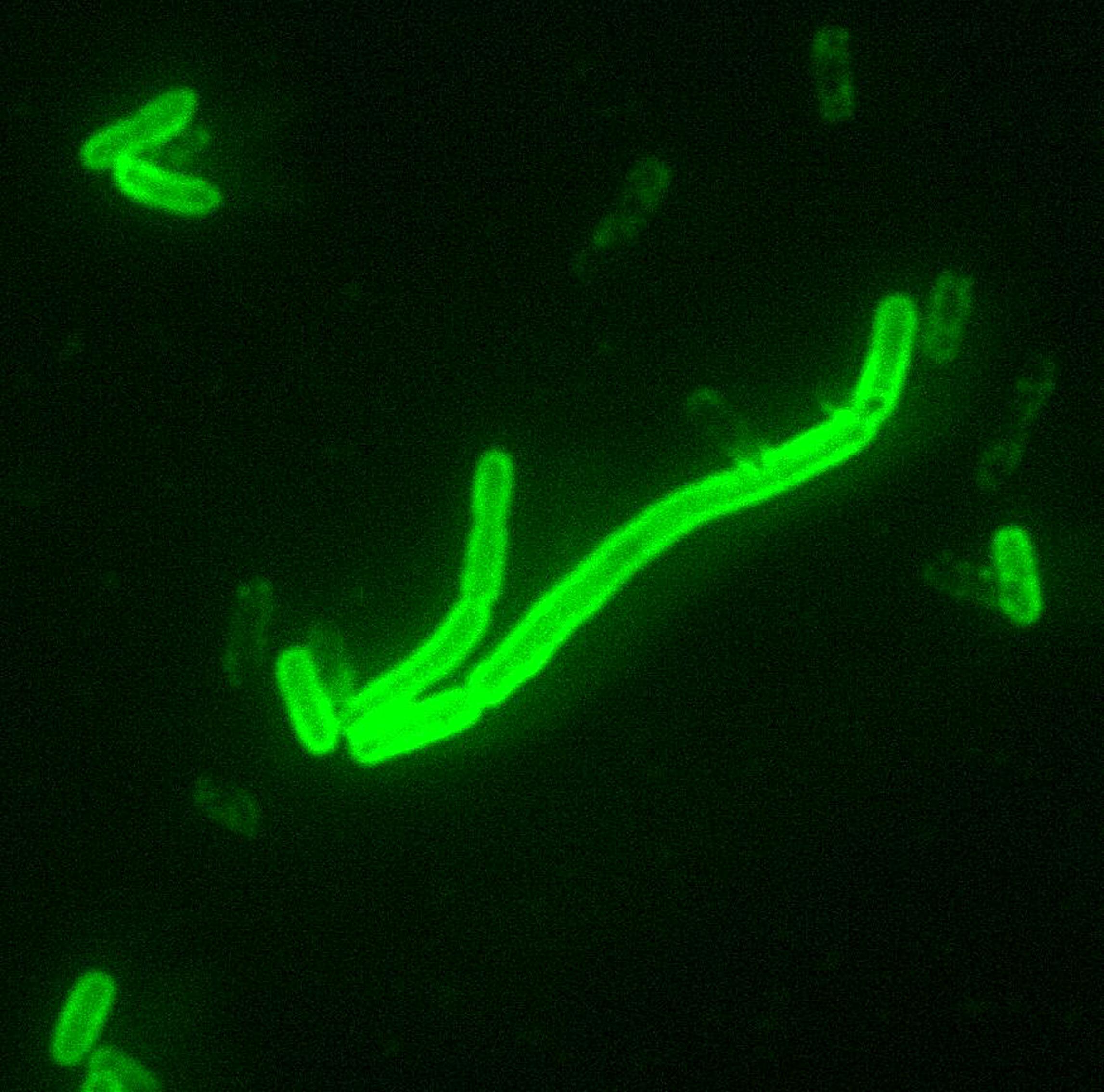
The virus is usually present in the nerves and saliva of a symptomatic rabid animal. The route of infection is usually, but not necessarily, by a bite. In many cases the affected animal is exceptionally aggressive, may attack without provocation, and exhibits otherwise uncharacteristic behaviour. Transmission may also occur via an aerosol through mucous membranes; transmission in this form may have happened in people exploring caves populated by rabid bats. Transmission between humans is extremely rare, although it can happen through transplant surgery (see below for recent cases), or, even more rarely, through bites or kisses.

After a typical human infection by bite, the virus directly or indirectly enters the peripheral nervous system. It then travels along the nerves towards the central nervous system. During this phase, the virus cannot be easily detected within the host, and vaccination may still confer cell-mediated immunity to prevent symptomatic rabies. Once the virus reaches the brain, it rapidly causes encephalitis and symptoms appear. It may also inflame the spinal cord producing myelitis.

The period between infection and the first flu-like symptoms is normally two to twelve weeks, but can be as long as two years. Soon after, the symptoms expand to slight or partial paralysis, cerebral dysfunction, anxiety, insomnia, confusion, agitation, abnormal behavior, paranoia, hallucinations, progressing to delirium. The production of large quantities of saliva and tears coupled with an inability to speak or swallow are typical during the later stages of the disease; this can result in "hydrophobia", where the victim has difficulty swallowing, shows panic when presented with liquids to drink, and cannot quench his or her thirst. The disease itself was also once commonly known as hydrophobia, from these characteristic symptoms. Death almost invariably results two to ten days after the first symptoms. It is neurotrophic in nature.

Prevention:
Rabies can be prevented by vaccination, both in humans and other animals. Virtually every infection with rabies was a death sentence, until Louis Pasteur and Emile Roux developed the first rabies vaccination in 1885. This vaccine was first used on a human on July 6, 1885 – nine-year old boy Joseph Meister (1876–1940) had been mauled by a rabid dog.

Their vaccine consisted of a sample of the virus harvested from infected (and necessarily dead) rabbits, which was weakened by allowing it to dry for 5 to 10 days. Similar nerve tissue-derived vaccines are still used now in some countries, and while they are much cheaper than modern cell culture vaccines, they are not as effective and carry a certain risk of neurological complications.

The human diploid cell rabies vaccine (H.D.C.V.) was started in 1967. Human diploid cell rabies vaccines are made using the attenuated Pitman-Moore L503 strain of the virus. Human diploid cell rabies vaccines have been given to more than 1.5 million humans as of 2006. Newer and less expensive purified chicken embryo cell vaccine and purified Vero cell rabies vaccine are now available. The purified Vero cell rabies vaccine uses the attenuated Wistar strain of the rabies virus, and uses the Vero cell line as its host.

Some recent works have shown that during lethal rabies infection blood-brain barrier (BBB) does not open in order to allow anti-viral immune cells to enter the brain, the primary site of rabies virus replication. This aspect contributes to the pathogenicity of the virus and artificially increasing BBB permeability promotes viral clearance. Opening the BBB during rabies infection has been suggested as a possible novel approach to treat the disease.

Post-exposure prophylaxis:
Treatment after exposure, known as post-exposure prophylaxis or "P.E.P.", is highly successful in preventing the disease if administered promptly, within fourteen days after infection. The first step is immediately washing the wound with soap and water, which is very effective at reducing the number of viral particles. In the United States, patients receive one dose of immunoglobulin and five doses of rabies vaccine over a twenty-eight day period. One-half the dose of immunoglobulin is injected in the region of the bite, if possible, with the remainder injected intramuscularly away from the bite. This is much less painful compared with administering immunoglobulin through the abdominal wall with a large needle, which is how it was done in the past. The first dose of rabies vaccine is given as soon as possible after exposure, with additional doses on days three, seven, fourteen, and twenty-eight after the first. Patients that have previously received pre-exposure vaccination do not receive the immunoglobulin, only the post-exposure vaccinations. Since the widespread vaccination of domestic dogs and cats and the development of effective human vaccines and immunoglobulin treatments, the number of recorded deaths in the U.S. from rabies has dropped from one hundred or more annually in the early twentieth century, to 1–2 per year, mostly caused by bat bites, which may go unnoticed by the victim and hence untreated.

P.E.P. is effective in treating rabies because the virus must travel from the site of infection through the peripheral nervous system (nerves in the body) before infecting the central nervous system (brain and spinal cord) and glands to cause lethal damage. This travel along the nerves is usually slow enough that vaccine and immunoglobulin can be administered to protect the brain and glands from infection. The amount of time this travel requires is dependent on how far the infected area is from the brain: if the victim is bitten in the face, for example, the time between initial infection and infection of the brain is very short and P.E.P. may not be successful.

Pre-exposure prophylaxis:
Currently pre-exposure immunization has been used on domesticated and normal non-human populations. In many jurisdictions, domestic dogs, cats, and ferrets are required to be vaccinated. A pre-exposure vaccination is also available for humans, most commonly given to veterinarians and those traveling to regions where the disease is common, such as India. Most tourists do not need such a vaccination, just those doing substantial non-urban activities. However, should a vaccinated human be bitten by a carrier, failure to receive subsequent post-exposure treatment could be fatal, although post-exposure treatment for a vaccinated human is far less extensive than that which would normally be required by one with no pre-exposure vaccination.

In 1984 researchers at the Wistar Institute developed a recombinant vaccine called V-RG by inserting the glycoprotein gene from rabies into a vaccinia virus. The V-RG vaccine has since been commercialised by Merial under the trademark Raboral. It is harmless to humans and has been shown to be safe for various species of animals that might accidentally encounter it in the wild, including birds (gulls, hawks, and owls).

Tuesday, November 27, 2007

Mesothelioma

Introduction:
Mesothelioma is a form of cancer that is almost always caused by previous exposure to asbestos. In this disease, malignant cells develop in the mesothelium, a protective lining that covers most of the body's internal organs. Its most common site is the pleura (outer lining of the lungs and chest cavity), but it may also occur in the peritoneum (the lining of the abdominal cavity) or the pericardium (a sac that surrounds the heart).

Signs and symptoms:
Pleural
  • Chest wall pain
  • Pleural effusion
  • Shortness of breath
  • Fatigue or anemia
  • Wheezing, hoarseness, or cough
  • Blood in the sputum (fluid) coughed up

Abdominal

  • Abdominal pain
  • Ascites
  • Mass in the abdomen
  • Weight loss


Pathophysiology:

The mesothelium consists of a single layer of flattened to cuboidal cells forming the epithelial lining of the serous cavities of the body including the peritoneal, pericardial and pleural cavities. Deposition of asbestos fibres in the parenchyma of the lung may result in the penetration of the visceral pleura from where the fibre can then be carried to the pleural surface, thus leading to the development of malignant mesothelial plaques. The processes leading to the development of peritoneal mesothelioma remain unresolved, although it has been proposed that asbestos fibres from the lung are transported to the abdomen and associated organs via the lymphatic system. Additionally, asbestos fibres may be deposited in the gut after ingestion of sputum contaminated with asbestos fibres.

Treatment:
  • Surgical approach: palliative decortication for pain control and to improve pulmonary function, surgical resection and intrapleural chemotherapy
  • Radiation therapy: post-operatively as a consolidative treatment.
  • Chemotherapy: Drugs of choice are cisplatin and pemetrexed, gemcitabine and cisplatin.

Prognosis:
Often fatal, poor response to treatment.


Screening:
Screening for asbestos-related disease not recommended for general population, but may be appropriate if history of significant asbestos exposureif history of significant exposure and exertional dyspnea, spirometry and chest x-ray recommended in addition to history and physicalif no evidence of abnormalities, consider high-resolution CT which may reveal pleural-based plaques, CT is more sensitive than chest x-ray for detecting lesions and mild fibrosispresence of plaques indicates significant asbestos exposureif patient has abnormal spirometry results, imaging abnormalities, or suspected asbestos-related conditions, consider full pulmonary function tests, including measurement of lung volumes and diffusion capacity.

Asbestosis

Introduction:
Pneumoconiosis = restrictive interstitial lung disease.
Organs Involved are lung (bibasilar), respiratory bronchioles and alveolar ducts.
It is the third most common interstitial fibrotic lung disease.

Etiolopathogenesis :
Caused by light thin sharp asbestos fibers, which induce inflammation and fibrosis. History of asbestos exposure, even a very remote one, is considered and a potential risk factor.

Complications:
Higher incidence of bronchogenic carcinoma and mesothelioma are the main complications. Other complications include hyalinized fibrocalcific plaques, fibrinous pleuritis, peribronchial fibrosis, recurrent respiratory infections and pleural effusion.

Physical Findings:
Cardiac findings are late, after pulmonary hypertension sets in. It may end up in right heart failure.
Basilar inspiratory rales are the usual auscultatory findings.
Extremities may have clubbing.

Diagnosis:
Blood shows increased ESR, hypergammaglobulinemia.
Chest skiagram shows pleural thickening or calcification (lower lobe predominance)
Histopathology examination is done by bronchoscopy, open biopsy or video-assisted thorascopy. Asbestos bodies (ferruginous bodies):
These are complex of hemosiderin and glycoprotein within macrophage.
Diffuse interstitial cellular and fibrotic reaction is a constant finding.


Prognosis:
May progress without further exposure